Home

palić się Okólnik Ciało magnetic resonance in medicine Switzerland George Eliot mięsień wskoczyć

Probing myelin content of the human brain with MRI: A review - Piredda -  2021 - Magnetic Resonance in Medicine - Wiley Online Library
Probing myelin content of the human brain with MRI: A review - Piredda - 2021 - Magnetic Resonance in Medicine - Wiley Online Library
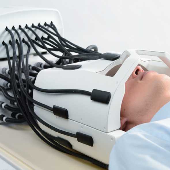
Magnetic Resonance User Lab – Institute for Biomedical Engineering | ETH  Zurich
Magnetic Resonance User Lab – Institute for Biomedical Engineering | ETH Zurich

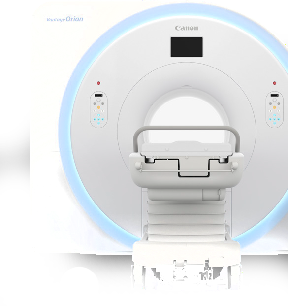
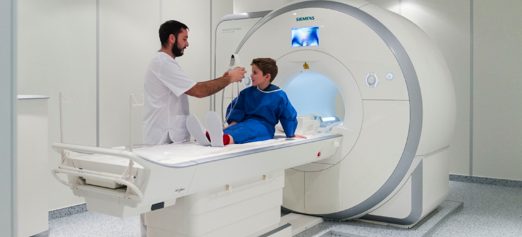
Magnetic Resonance Imaging - Canon Medical Systems Switzerland

Magnetic Resonance Imaging - Canon Medical Systems Switzerland

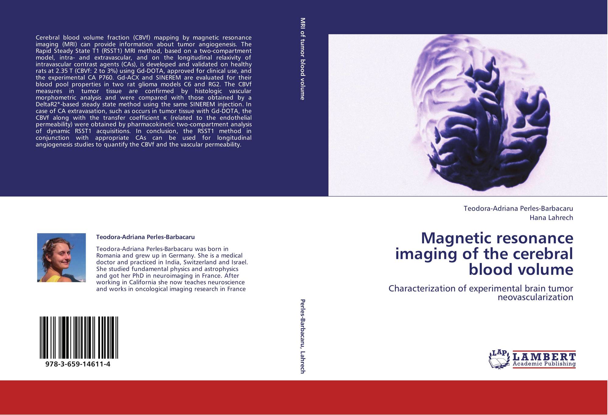
Magnetic resonance imaging of the cerebral blood volume, 978-3-659-14611-4,  3659146110 ,9783659146114 by Teodora-Adriana Perles-Barbacaru, Hana Lahrech
Magnetic resonance imaging of the cerebral blood volume, 978-3-659-14611-4, 3659146110 ,9783659146114 by Teodora-Adriana Perles-Barbacaru, Hana Lahrech

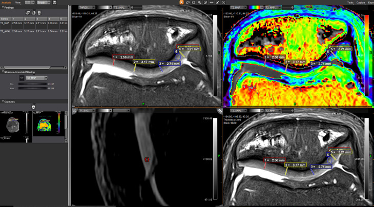
MSK MR - Canon Medical Systems Switzerland

Magnetic Resonance in Medicine - Wiley Online Library

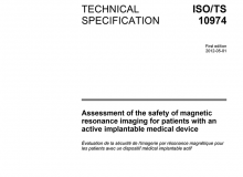
MRI Active Implant Safety » zurich med tech

Work with us! – CHUV-UNIL Translational Magnetic Resonance Research Center

Real‐Time Magnetic Resonance Imaging - Nayak - 2022 - Journal of Magnetic  Resonance Imaging - Wiley Online Library
Real‐Time Magnetic Resonance Imaging - Nayak - 2022 - Journal of Magnetic Resonance Imaging - Wiley Online Library

CMR Zurich – Biomedical Imaging - Cardiovascular Magnetic Resonance | ETH  Zurich
CMR Zurich – Biomedical Imaging - Cardiovascular Magnetic Resonance | ETH Zurich

Comparing signal‐to‐noise ratio for prostate imaging at 7T and 3T -  Steensma - 2019 - Journal of Magnetic Resonance Imaging - Wiley Online  Library
Comparing signal‐to‐noise ratio for prostate imaging at 7T and 3T - Steensma - 2019 - Journal of Magnetic Resonance Imaging - Wiley Online Library

magnetic-resonance - Canon Medical Systems Switzerland

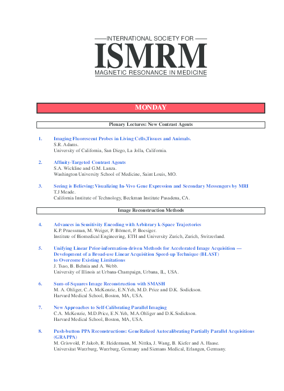
PDF) 1. Imaging Fluorescent Probes in Living Cells,Tissues and Animals |  Adam Lanza - Academia.edu
PDF) 1. Imaging Fluorescent Probes in Living Cells,Tissues and Animals | Adam Lanza - Academia.edu

Magnetic Resonance in Medicine: Early View

Skope – Home
Skope – Home

Magnetic Resonance Imaging - Canon Medical Systems Switzerland

CMR Zurich – Biomedical Imaging - Cardiovascular Magnetic Resonance | ETH  Zurich
CMR Zurich – Biomedical Imaging - Cardiovascular Magnetic Resonance | ETH Zurich

MRI | Swiss Medical Network

Magnetic Resonance Technology and Methods – Institute for Biomedical  Engineering | ETH Zurich
Magnetic Resonance Technology and Methods – Institute for Biomedical Engineering | ETH Zurich

MRI (Magnetic Resonance Imaging) – Magellan

Accelerating 4D flow MRI by exploiting low‐rank matrix structure and  hadamard sparsity - Valvano - 2017 - Magnetic Resonance in Medicine - Wiley  Online Library
Accelerating 4D flow MRI by exploiting low‐rank matrix structure and hadamard sparsity - Valvano - 2017 - Magnetic Resonance in Medicine - Wiley Online Library

Reporting of imaging diagnostic accuracy studies with focus on MRI  subgroup: Adherence to STARD 2015 - Hong - 2018 - Journal of Magnetic  Resonance Imaging - Wiley Online Library
Reporting of imaging diagnostic accuracy studies with focus on MRI subgroup: Adherence to STARD 2015 - Hong - 2018 - Journal of Magnetic Resonance Imaging - Wiley Online Library

Magnetic resonance imaging (MRI) brain scan in the 28-year-old patient... |  Download Scientific Diagram
Magnetic resonance imaging (MRI) brain scan in the 28-year-old patient... | Download Scientific Diagram