Home

Robić Mikroprocesor Mglisty protein 3d structure analysis Różnorodność Las indyk

High-res 3D structure of proteins inside living eukaryotic cells determined

Sequence co-evolution gives 3D contacts and structures of protein complexes  | eLife
Sequence co-evolution gives 3D contacts and structures of protein complexes | eLife

Predicted 3D structures for three representative proteins. Visual... |  Download Scientific Diagram
Predicted 3D structures for three representative proteins. Visual... | Download Scientific Diagram

Protein Structure Analysis Workflow | DNASTAR

Network analysis of protein structure for 1CRN (chain A). (A) Network... |  Download Scientific Diagram
Network analysis of protein structure for 1CRN (chain A). (A) Network... | Download Scientific Diagram
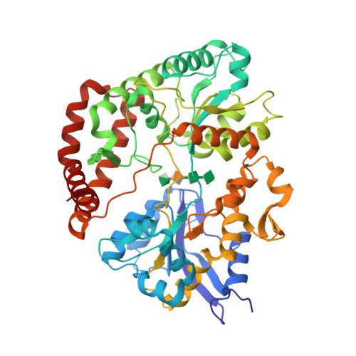
Protein 3D-structure analysis

RCSB PDB - 1J1N: Structure Analysis of AlgQ2, A  Macromolecule(Alginate)-Binding Periplasmic Protein Of Sphingomonas Sp.  A1., Complexed with an Alginate Tetrasaccharide
RCSB PDB - 1J1N: Structure Analysis of AlgQ2, A Macromolecule(Alginate)-Binding Periplasmic Protein Of Sphingomonas Sp. A1., Complexed with an Alginate Tetrasaccharide

Molecular-Recogunition Structure Analysis Team | Molecular Profiling  Research Center for Drug Discovery
Molecular-Recogunition Structure Analysis Team | Molecular Profiling Research Center for Drug Discovery

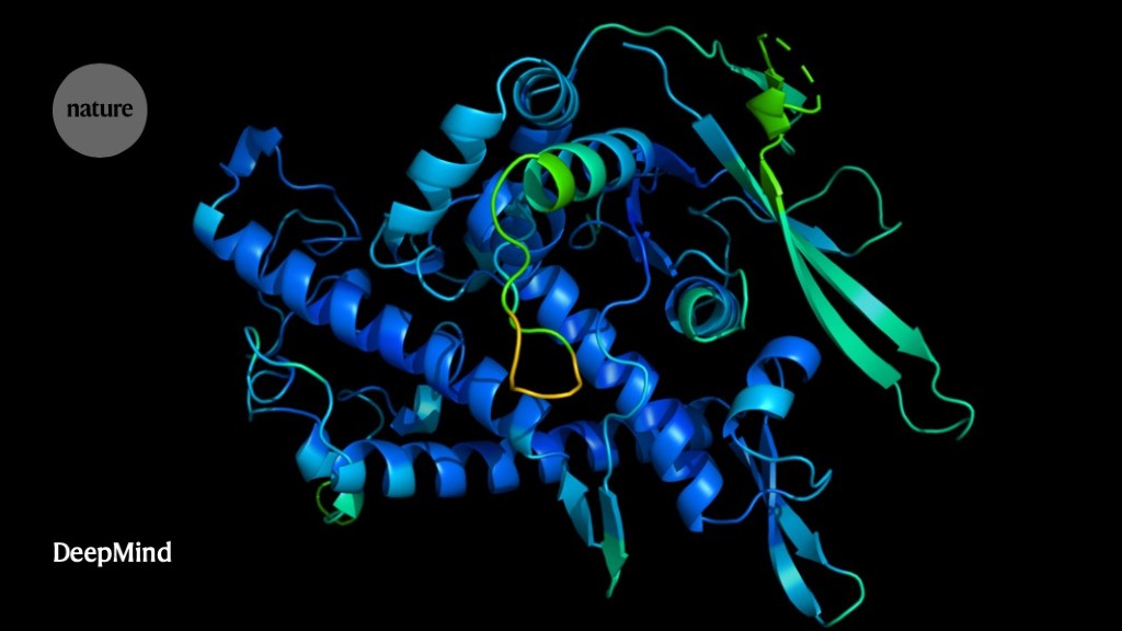
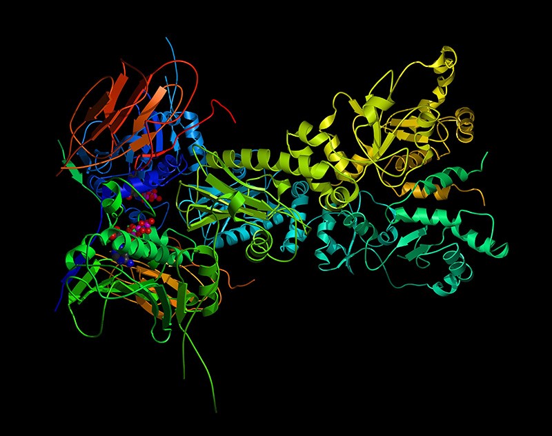
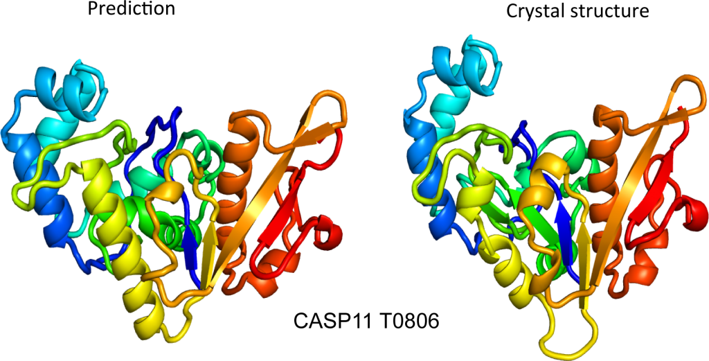
It will change everything': DeepMind's AI makes gigantic leap in solving protein  structures
It will change everything': DeepMind's AI makes gigantic leap in solving protein structures

Protein Analysis Techniques Explained - ATA Scientific

Go group : Research Area

2 Protein structural analysis of selected sequences from each gene... |  Download Scientific Diagram
2 Protein structural analysis of selected sequences from each gene... | Download Scientific Diagram

Functional characterization of 3D protein structures informed by human  genetic diversity | PNAS
Functional characterization of 3D protein structures informed by human genetic diversity | PNAS

Structure analysis of epitopes. 3D structures of protein targets with... |  Download Scientific Diagram
Structure analysis of epitopes. 3D structures of protein targets with... | Download Scientific Diagram

Functional characterization of 3D protein structures informed by human  genetic diversity | PNAS
Functional characterization of 3D protein structures informed by human genetic diversity | PNAS

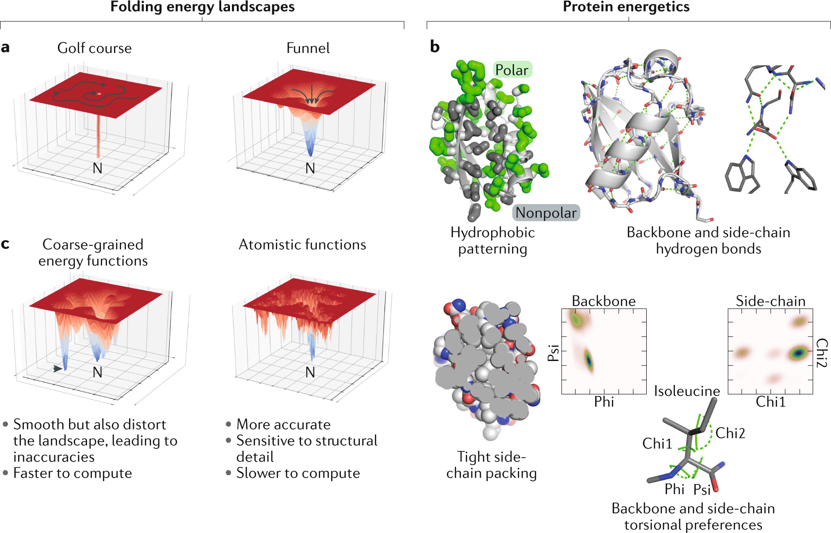
Advances in protein structure prediction and design | Nature Reviews  Molecular Cell Biology
Advances in protein structure prediction and design | Nature Reviews Molecular Cell Biology

Structure alignment of four PDZ proteins and results of position... |  Download Scientific Diagram
Structure alignment of four PDZ proteins and results of position... | Download Scientific Diagram

AI protein-folding algorithms solve structures faster than ever

Functional domain, 3D structure and structural analysis of zCDKL1. (A)... |  Download Scientific Diagram
Functional domain, 3D structure and structural analysis of zCDKL1. (A)... | Download Scientific Diagram

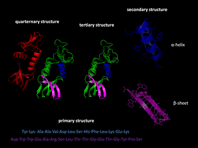
Protein structure - Wikipedia

3D protein structure and functional domain analysis of Gαq (GNAQ) and... |  Download Scientific Diagram
3D protein structure and functional domain analysis of Gαq (GNAQ) and... | Download Scientific Diagram

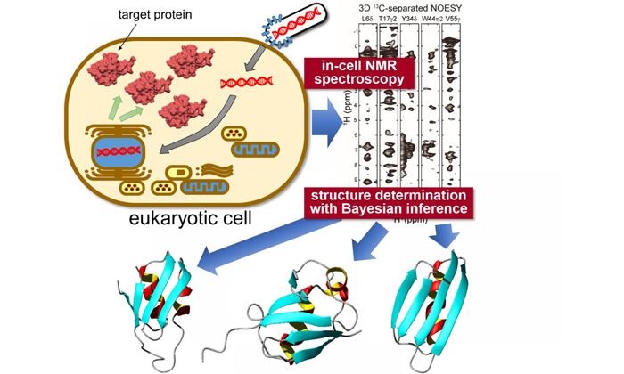
Protein Structural Analysis

November 2015 – Institute for Protein Design

Statistical prediction of protein structural, localization and functional  properties by the analysis of its fragment mass distributions after  proteolytic cleavage | Scientific Reports
Statistical prediction of protein structural, localization and functional properties by the analysis of its fragment mass distributions after proteolytic cleavage | Scientific Reports